
"Skeletons are shown within the flesh." (Image by Albert Londe.)
It was early in 1896 that X-rays became an important part of medical procedures, though researchers began working on its development as far back as 1875. German physics professor Wilhelm Röentgen ultimately got the credit for perfecting the process. In the February 10, 1896 Brooklyn Daily Eagle, the new invention is heralded for the major advancement it was. An excerpt:
“Until some man invents a camera that will take a picture around a corner the new utilization of the Cathode or Roentgen rays will suffice for wonder. In this process, as everyone knows by this time, objects that we have supposed to be opaque have been pierced by light so that objects within them have shown in shadowy mass on the photographic plate. A razor had been photographed inside of its case, skeletons are shown within the flesh and things have been revealed that were covered with black paper, wood, horn, rubber and thin plates of metal. Indeed, we begin to inquire if there is such a thing as opacity, now. All this is unexpected and curious, but to men most important for one thing: It opens the human body to examination. If something is wrong inside of one it may not be necessary to cut him open to find what. If the something is known it will save unnecessary cutting.
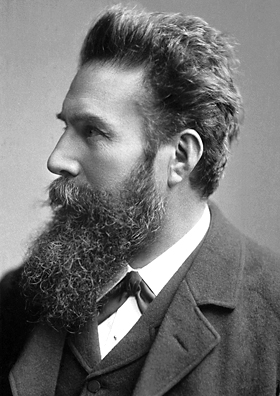
Wilhelm Röentgen, father of the X-ray. (Image by the Nobel foundation.)
To one who has never seen an operation by surgery, or has given small attention to such matters, it may seem as if it were easy to find the lodging place of a bullet or broken knife point in the flesh, but often the finding of a needle in a bundle of hay is an easy task compared with it. The hay, at all events, contains no nerves, no quivering muscles, no tough cartilage and tendon, no resisting bones: one does not have to be careful how he explores this way and that lest he cut an important nerve or sever an artery or tap a vein, And sometimes, after probing and reaching and cutting for half an hour the work is found to be too difficult and dangerous to continue; then the patient is made as easy as circumstances allow and told to resign himself to wearing the bullet or the knife point for the rest of his days.
By use of the X rays the bullet can be made to declare itself to the sight and the surgeon can go straight to it with his scalpel, and if the ball is found to lie in too close contact to an artery it can be left to encyst. In case of a compound fracture pieces of bone that may be driven into adjacent muscle may be promptly located and removed or replaced. Perhaps a higher sensitization can be obtained so that relatively opaque tumors, cancers, fungoid growths, ossifications or chalk deposits may be indicated in the picture and the surgeon will then be guided as to the mode of procedure. Appendicitis may be resolved into a case for surgical or for medical treatment according to what it shows of the degree of induration or suppuration or the presence of foreign and irritating bodies. Of all the recent advancement in surgery this of the employment of the cathode ray promises the most benefit.”
Tags: Wilhelm Röentgen